
No antibody? You may not have to build the assay from scratch

The protocol you already have is also the design brief
Many protein assays and immunoassays are built around antibodies. Sometimes the antibody is the part you cannot get. The target is too similar across species to raise a useful immune response. Or it holds the relevant shape only in a membrane and loses it during purification. Or antibodies exist, but no validated pair does.
The format itself is ordinary. A sandwich ELISA, a bead assay, a lateral-flow strip. Thousands of related assays have been run and detailed protocols exist – just not for your target.
An aptamer is one route around the missing antibody: a short DNA or RNA sequence that folds into a structure capable of binding a target. It is not a small antibody, and it cannot be dropped into an antibody protocol unchanged. But the protocol still carries most of what it cost to develop. And it carries more than the assay.
Where the design inputs come from
Structure-based design settles the target region, the attachment terminus, the label position and the working conditions before anything is synthesised. Each still has to be confirmed at the bench, since a predicted site is a hypothesis, but the first attempt has a reason behind it. Variants also stop being a bottleneck: two label positions and three spacer lengths are one synthesis order, not a separate conjugation and clean-up campaign for every configuration.
Almost everything a design run needs is written in the antibody protocol you are already holding.
| In the protocol | Becomes a design input |
|---|---|
| Binding buffer and sample diluent | The ionic conditions the fold is built for |
| Incubation temperature | The temperature the structure has to hold at |
| Surface chemistry (streptavidin plate, gold, amine slide) | Which anchor chemistry the construct needs |
| Capture or detection role | Which end stays free |
| Reporter chemistry | Which modification the sequence has to carry |
| Intended measuring range and decision point | The affinity and kinetic regime to aim for |
| Cross-reactants already ruled out | Off-targets and the cross-signal each is allowed |
| Sample matrix | Nuclease exposure, ionic conditions |
Two things the protocol does not supply. One is the target structure itself, along with the decision about which conformational state, which domain boundaries and which modification state it represents. Design against a form that does not occur in your samples and you have made the same mistake as selecting against the wrong recombinant. The other is the assay's actual analytical range and cutoff, which come out of validation. The protocol gives you the range you are aiming at. What you end up reporting comes out of the work.
Everything else in the table is already written down. Collecting the brief is transcription; what a design run does with it is not.
What the protocol also constrains
The same document narrows the assay itself. The architecture, sandwich or competitive, and the order of the steps. The detection and amplification chemistry. The plate, bead or reader configuration. The washing scheme and starting windows for incubation times. The dynamic range the platform supports. A candidate sample workflow, if the matrix is comparable. And the failure modes somebody has already run into.
These narrow the search. None of them is a result you can claim for your own assay.
What is left
Four questions belong to the binder: how much of it goes in, how it folds, how it attaches, and how you show that it works. Each has its own experiment.
How much goes in
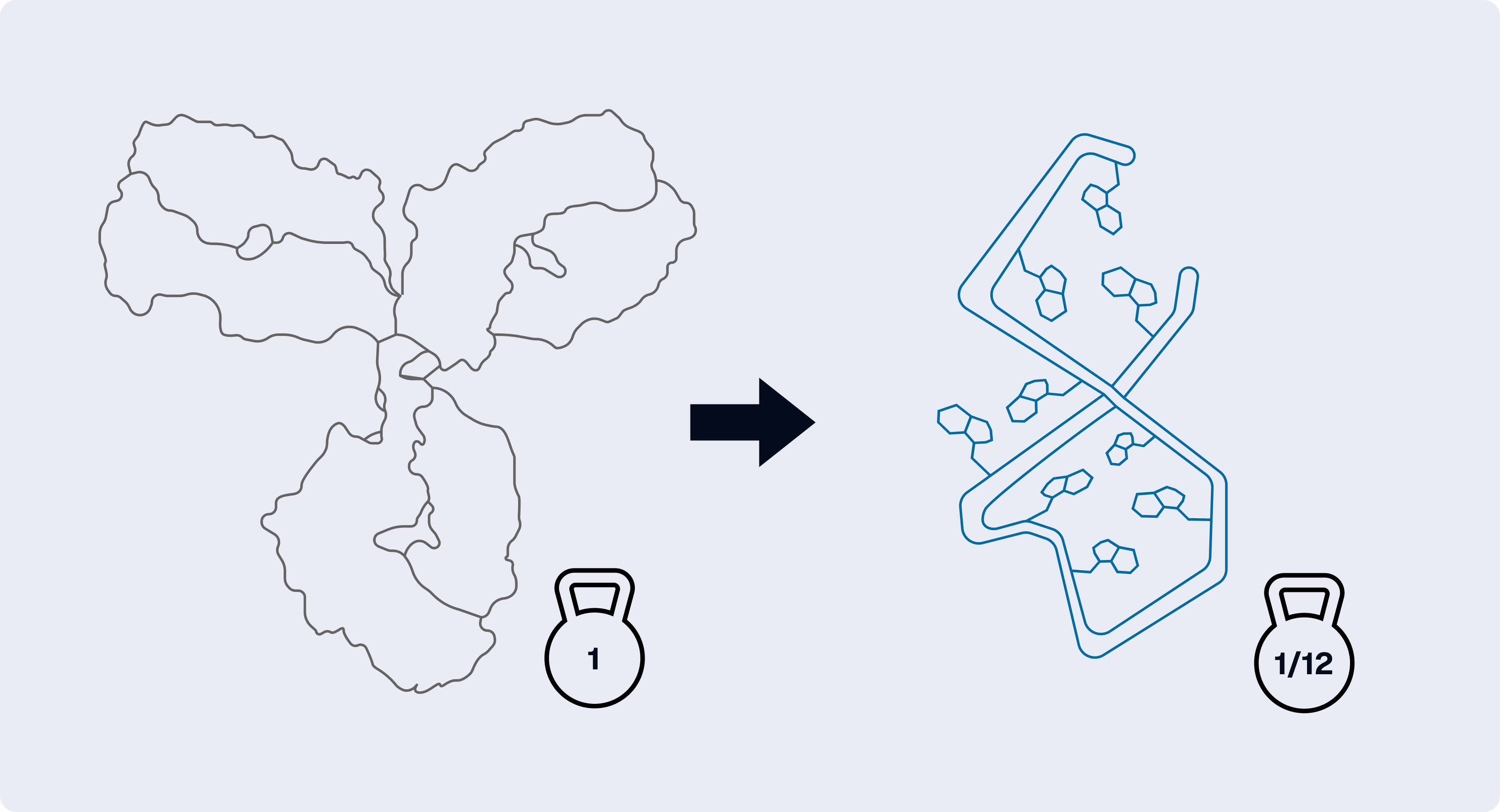
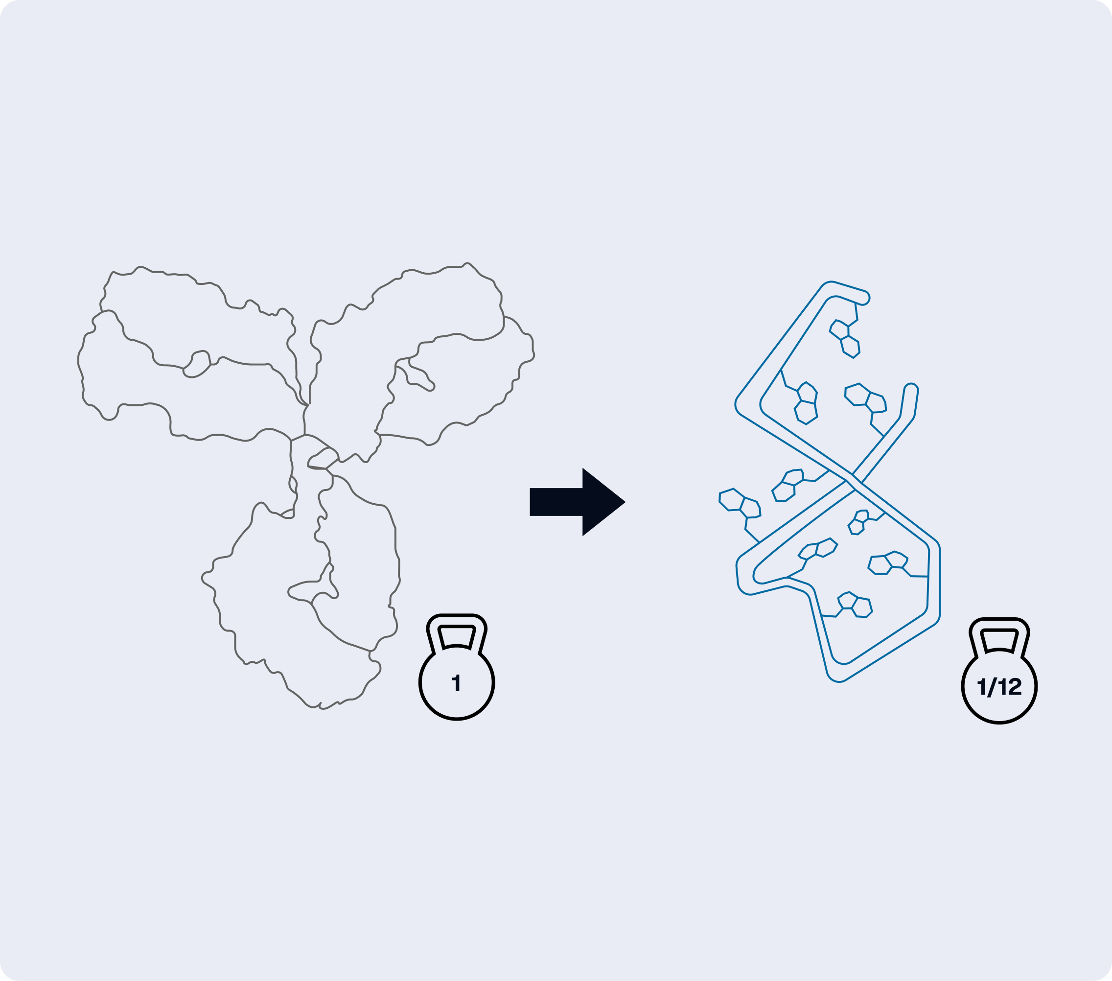
A typical IgG is about 150 kDa. A forty-nucleotide DNA aptamer is roughly 12–13 kDa. At the same mass concentration, the aptamer solution holds about twelve times as many molecules, so a µg/mL figure copied across from an antibody protocol lands an order of magnitude off.
Scale by the ratio of the masses instead:
A protocol calling for 1 µg/mL of a 150 kDa antibody gives about 83 ng/mL for a 12.5 kDa aptamer. Use the exact mass from the manufacturer, labels and linkers included: on a short sequence a label can shift the mass by several hundred daltons, far more than base composition does.
That is a starting point for the first titration, not a working concentration. Test a quarter, a half, one, two and four times it. For an immobilised capture reagent it is weaker still, because what counts there is how many accessible sites end up on the surface. And do not correct for the antibody's two binding sites with a factor of two: the fraction of sites still accessible on a surface is unknown, as is the fraction of aptamers correctly folded. Titration settles it; arithmetic does not.
How it folds
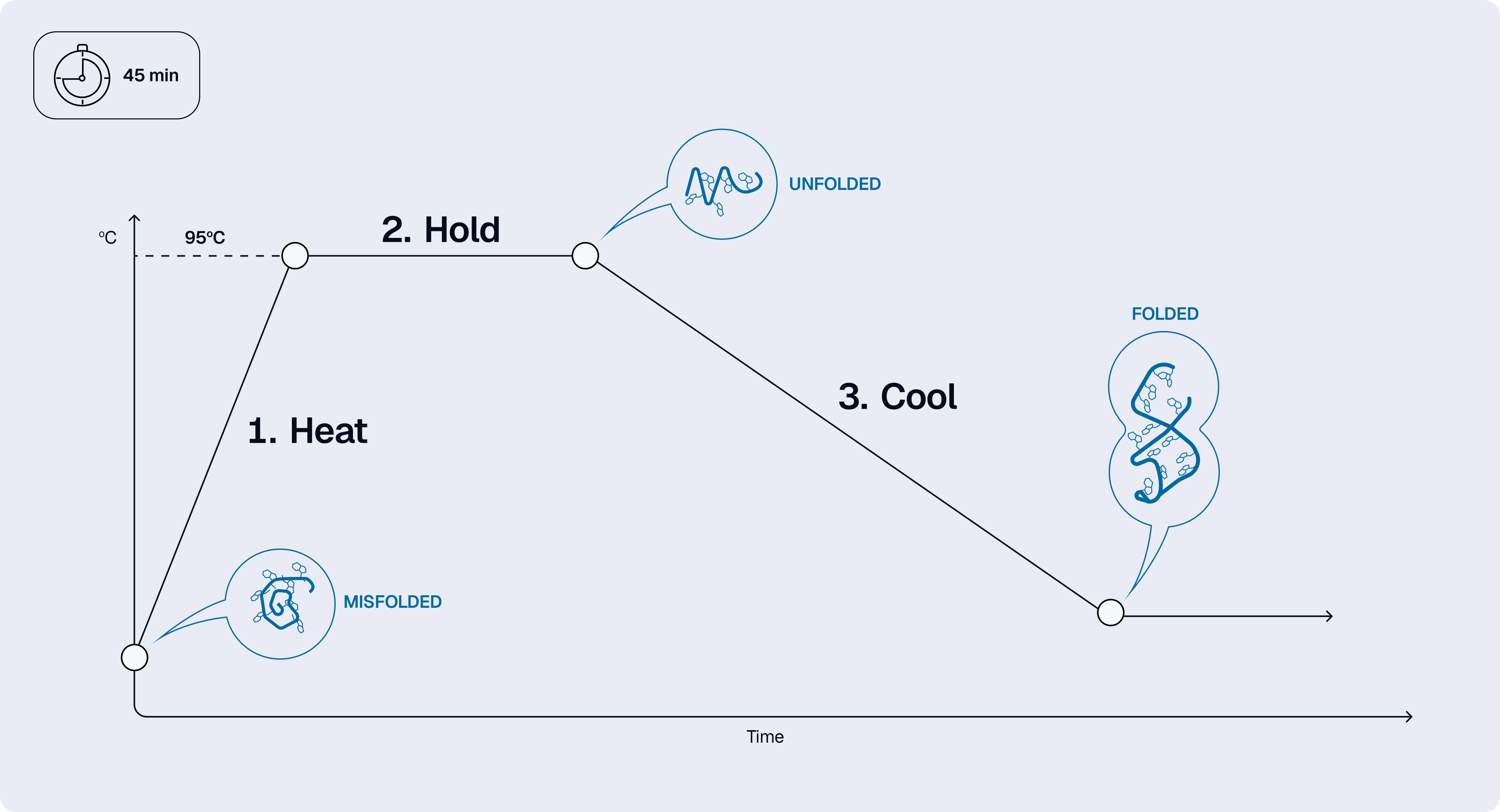
An aptamer has to be in the right shape before it can bind, and it does not always arrive that way. Some sequences hold their structure; others only form it on contact with the target. Which shapes are available depends on sequence, buffer, ions, temperature and concentration.
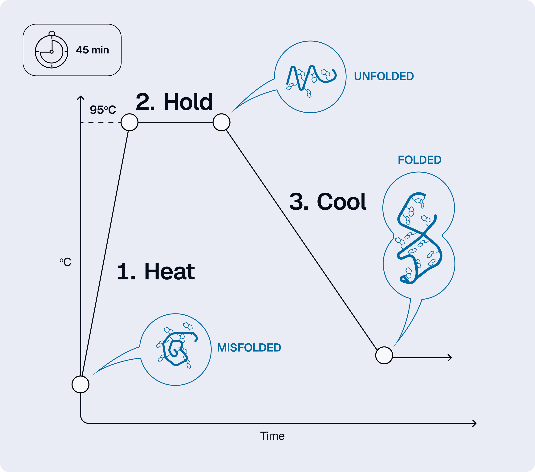
So refold the reagent before use, on the conditions supplied with it. For unmodified DNA that usually means heating to around 95 °C, holding a few minutes, then cooling to the working temperature and letting it settle there – under an hour on a heat block. RNA, modified backbones and some labels have their own limits, and what came with the sequence wins.
Dilute before heating: that reduces pairing between separate strands, which depends on concentration, while an intramolecular fold does not. Then record what you ran – temperature, duration, cooling rate and the concentration during heating. ‘Heated and cooled before use’ does not let anyone repeat the experiment, including you.
Buffer is the one line of the brief you read instead of copying. An antibody protocol's buffers were chosen for a protein: EDTA in a diluent, or a chelator in a blocking solution, is harmless to an antibody and can strip the divalent cations an aptamer's structure depends on. That is why it sits in the first row of the table. Hand the buffer to the design run and the sequence is designed and scored for folding and binding under those conditions.
How it attaches
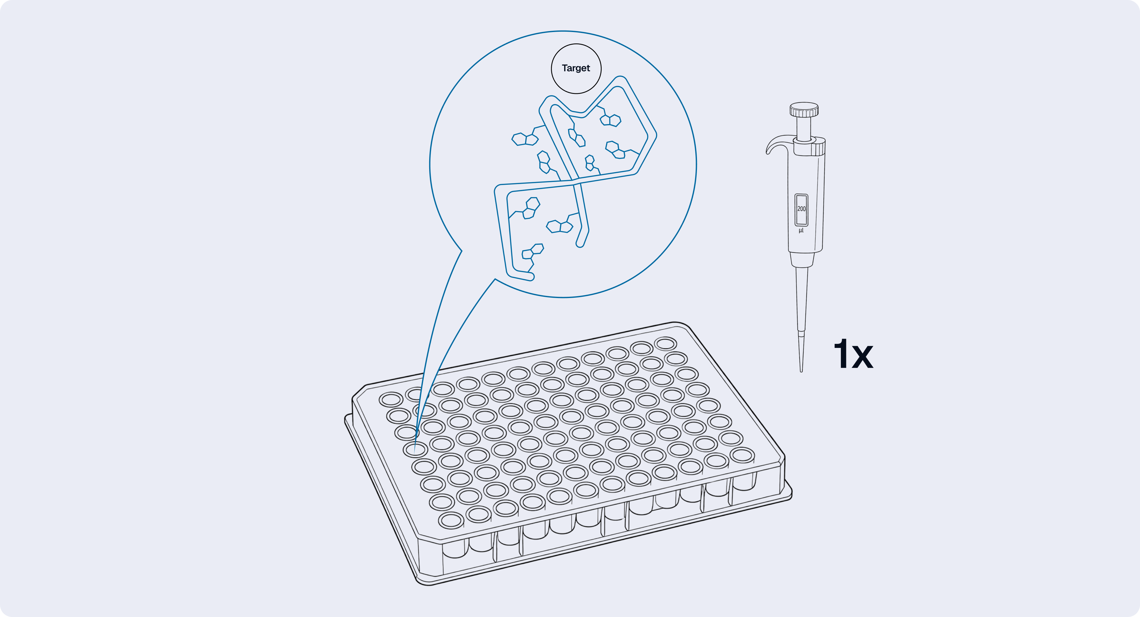
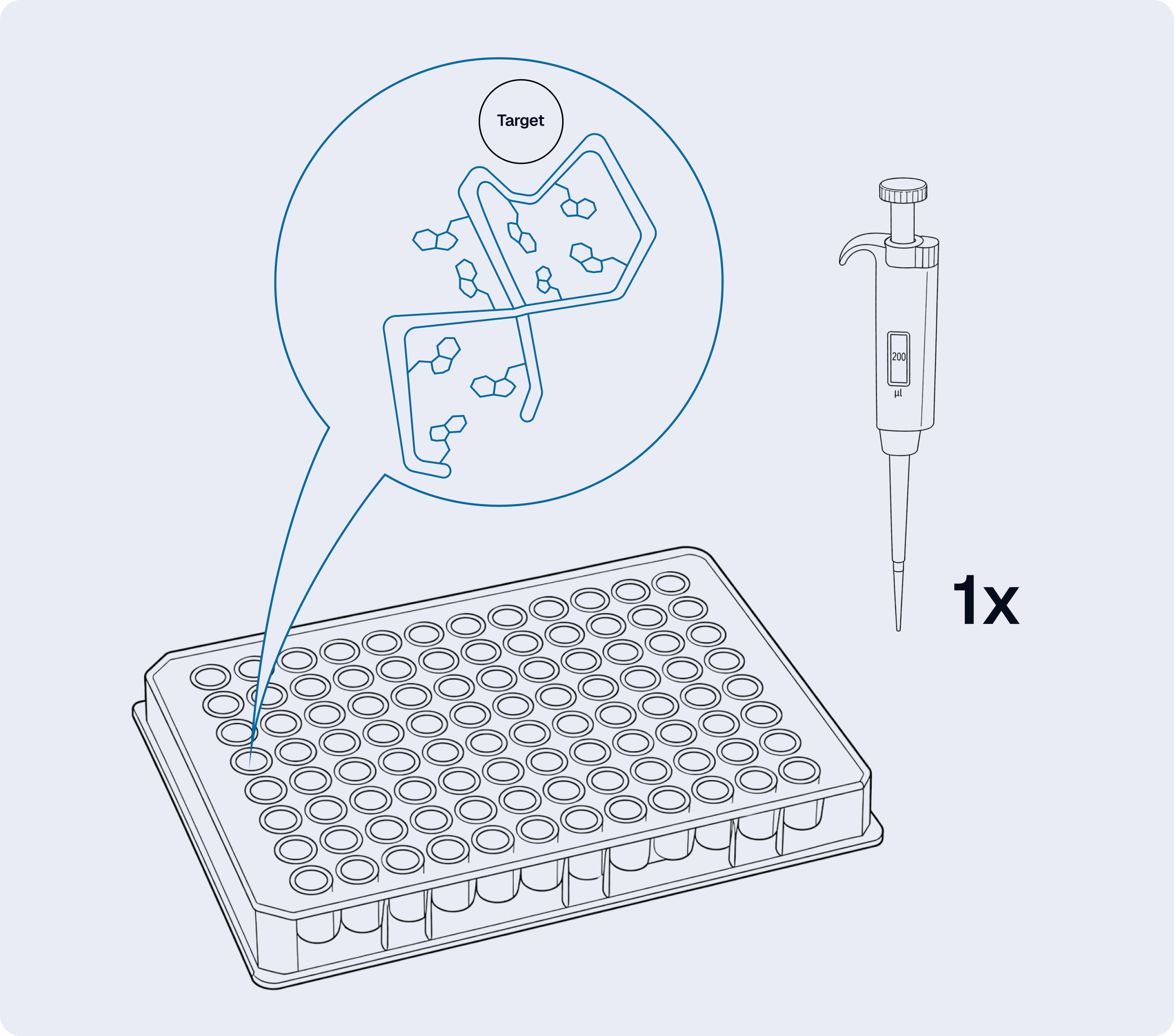
An aptamer can bind well in solution and lose most of its activity once immobilised. That does not by itself mean the sequence is unsuitable. How it sits on the surface is a separate question.
Some antibodies stay active after passive adsorption because a fraction of them land in a usable orientation. A short oligonucleotide has less tolerance: pressed against a surface, it may not be able to form the structure at all. A defined terminal anchor and a spacer usually help, and three variables carry the weight: which end holds the anchor, how long the spacer is, and how densely the molecules sit. Dense packing raises capacity and lowers the fraction that folds and binds correctly. Sparse packing preserves activity and gives too little signal.
A small screen answers this: two attachment configurations, two or three loading levels, and a second blocking buffer alongside the inherited one.
Before any of that, confirm binding in solution, in a buffer close to the assay's and with the reagent in its final chemical form. A result from the unmodified sequence does not transfer: the same label on the opposite end can leave binding untouched or abolish it.
For serum, plasma or lysate, add one stability experiment early. Incubate the final reagent in the intended matrix, at the planned dilution, for the full assay duration, then measure what remains and whether it still binds. Unmodified RNA is often degraded quickly. DNA holds up better, but not automatically.
Where the format needs two binders, build one layer at a time. If a suitable antibody exists for one side, keep it. The hybrid inherits a validated surface or a validated detection chain, and only one interface is new. Its epitope is another line in the design brief: aim the aptamer away from it.
If neither binder exists, test each aptamer alone, then run the experiment that shows both can hold the target at once. Independent affinity measurements are not evidence of pair compatibility, and a predicted non-overlapping site improves the odds without removing the experiment.
How you show that it works
If an antibody assay exists, run it alongside yours – same samples, same session, same hands – and decide in advance what agreement should mean. Do not expect identical signals: the reagents differ in size, kinetics and label geometry. Discordant samples get a predefined investigation, not a post hoc explanation: epitope accessibility, isoforms, modification state, matrix effects, kinetics, and error in the comparator itself.
More often there is nothing to compare against. Then the evidence has to converge from several directions.
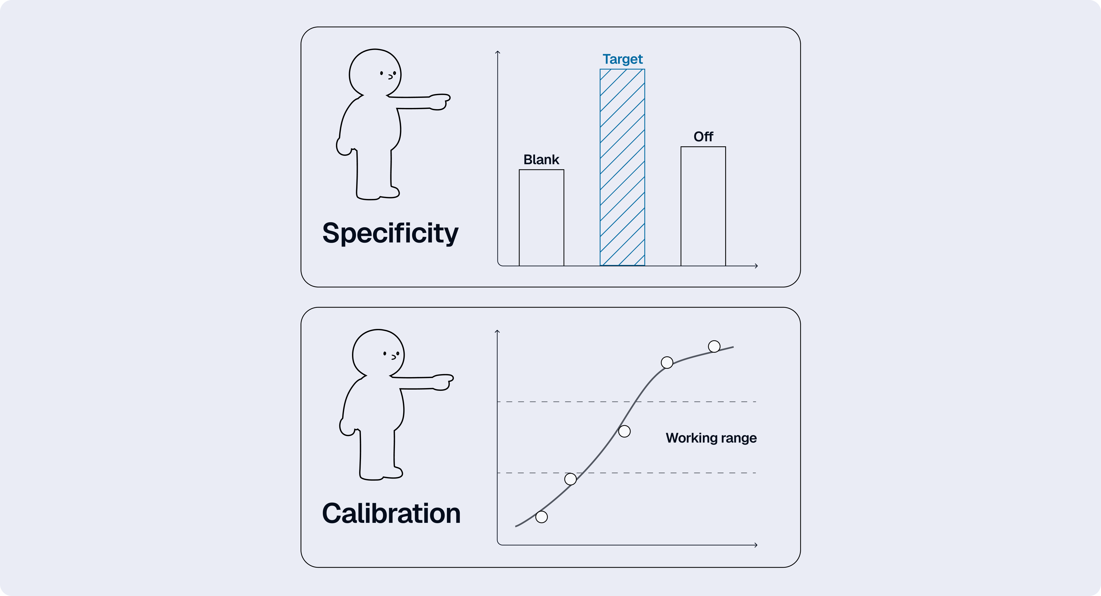
Recovery of a known amount of target added to real samples, at low, middle and high concentration. Dilutional linearity: a diluted sample should read the expected fraction. An independent measurement on a subset, where the analyte offers one: mass spectrometry, an activity assay.
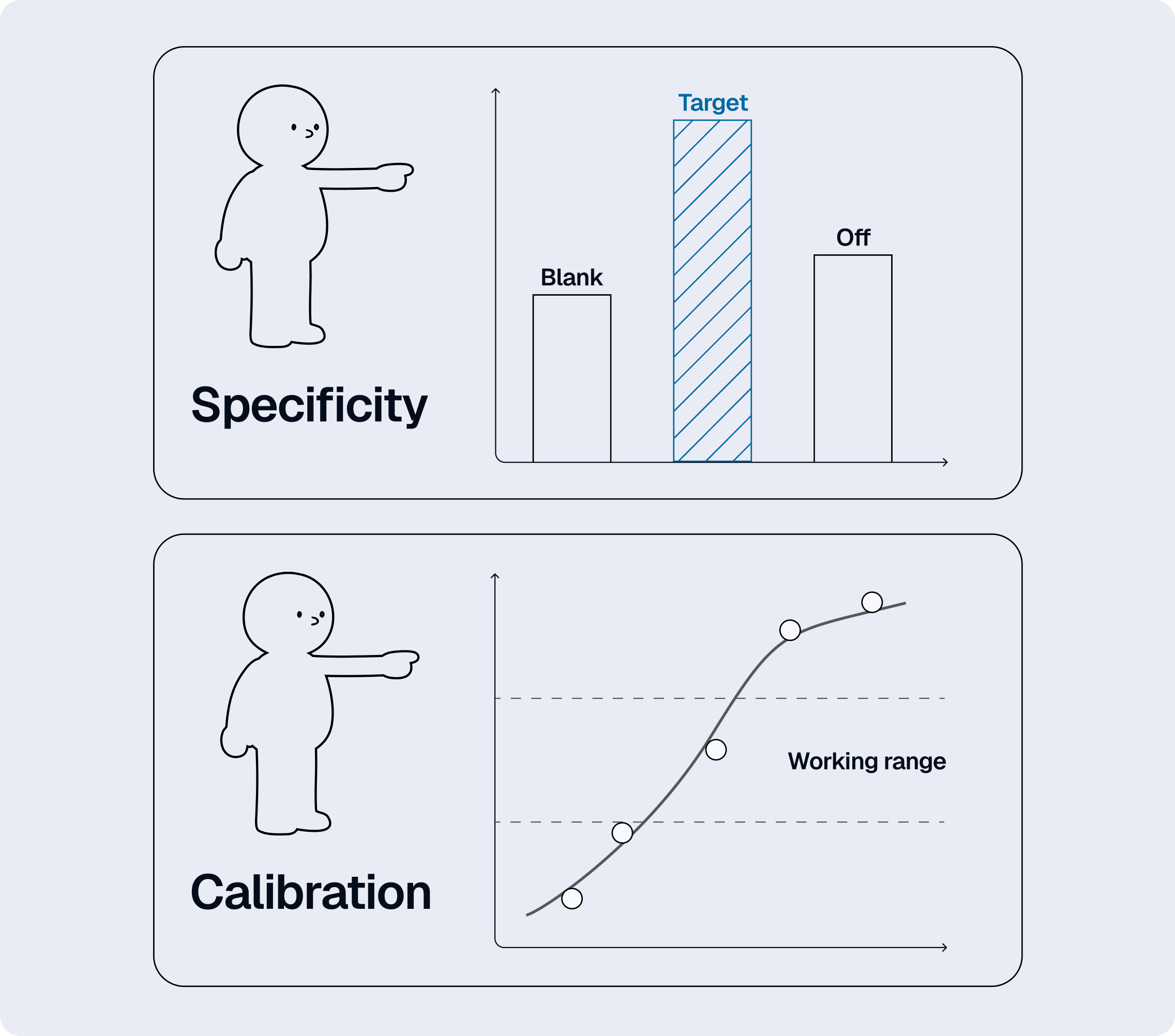
And a sequence control, which does more work here than anywhere else: it is what tells you the signal came from the sequence, and not from a charged polymer settling on the plate. Use one of similar length, chemistry, label and attachment, and confirm experimentally that it does not bind. A scrambled version of your own sequence is a candidate control, not a control: scrambling reorders the bases without guaranteeing an inactive structure. Competition with free target, and an unrelated protein of comparable size and charge, test the same thing from the other side.
Then evaluate the finished assay like any other method: usable calibration range, background, precision within and between runs, recovery, selectivity against related molecules and matrix components, robustness to small changes in timing and preparation. Set the acceptance criteria before looking at the results. With no established comparator that matters more than usual, because criteria written afterwards define success around whatever the assay happened to do.
The assay comes first
The usual order runs the other way. You obtain a binder, then work out what it needs – which buffer keeps it folded, which end tolerates a label, how much of it a well will hold – and the assay gets adjusted around the answers.
An antibody protocol lets you invert that. The requirements are already written down: buffer, temperature, anchor chemistry, label position, the range binding has to stay responsive across, the molecules that have to stay unbound. Hand them over before synthesis and the sequence is built against a specification instead of being discovered and then accommodated.
Four questions still belong to the bench: how much reagent goes in, how it folds, how it attaches, and how you show that the number means something. None of that guarantees a particular aptamer will work. It does mean that when one fails, you know which of the four to look at first, and which experiment to repeat.
© 2026 Xelari Inc. All rights reserved.
