
How to choose the right binding site for your aptamer
Where the aptamer binds decides what it can be used for
An aptamer can hit its target at single-digit nanomolar and still be useless. The site it found gets buried when the protein joins its complex. Or it sits under a glycan. Or it belongs to a domain that never appears in the fragment of the protein actually circulating in your samples. Or the binding is tight and reproducible and it changes nothing about the process you wanted to control.
Choosing the target protein is only the first decision. The next one is where on that protein the aptamer should bind, and what should happen when it does.
The previous article, The proteome doesn't have a coverage gap – it has a coverage bias, described three reasons proteins fall outside large measurement panels: they are hard to present in native form, they resemble their relatives too closely, or they exist as several biologically distinct forms that assays cannot separate. All three come back at the design stage, only now as questions about which patch of surface to aim at.
A protein offers more than one place to bind
Structures are full of plausible sites. Some are exposed and chemically inviting. Some are functionally decisive. The overlap between those two sets is smaller than you would like.
A convenient patch will give you a stable complex and no biological effect. The residue that actually matters often sits at the bottom of a cleft shaped for a small substrate rather than for a folded oligonucleotide. And a surface that looks wide open in a crystal structure of the isolated domain can turn out to be the interface the protein uses to dimerise.
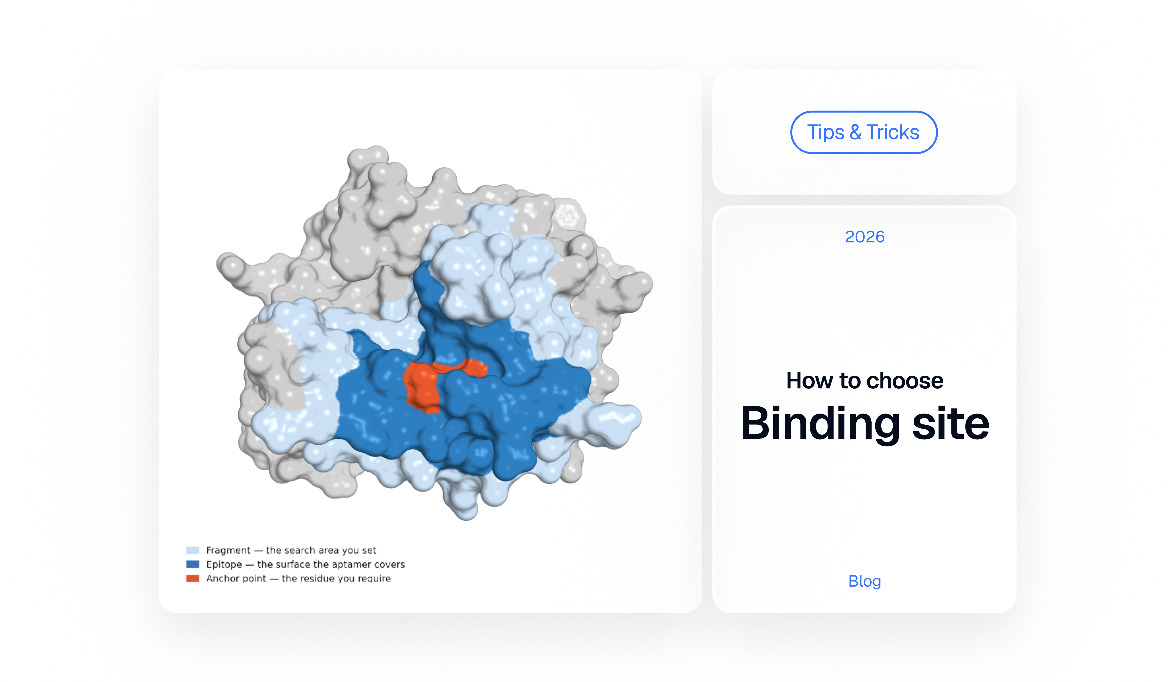
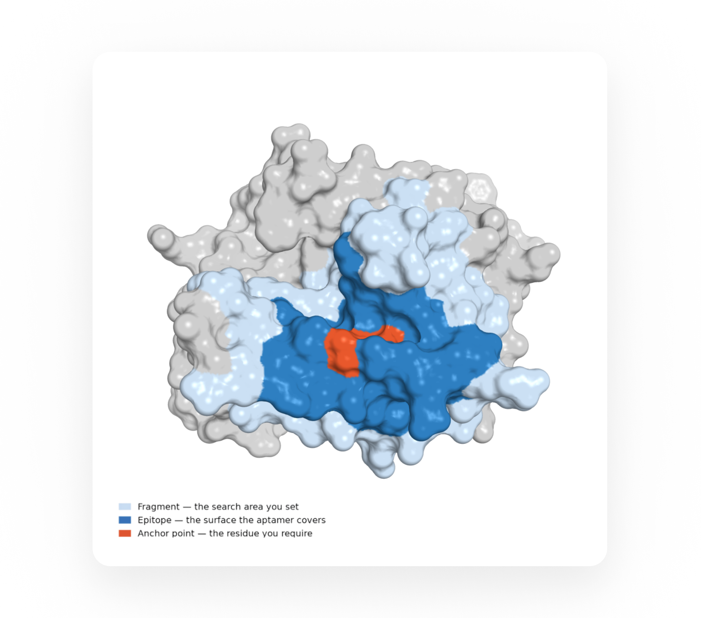
This is where three terms are worth separating: the fragment, the anchor point and the epitope.
Think of the protein surface as a city. The fragment is the district you send the search into. The anchor point is a particular house in that district: you give the address and require that the design include it. The epitope is the block the aptamer ends up covering – that house plus whichever neighbours come with it.
The fragment sets the search area
The fragment marks the structural region where design should look for a productive binding mode.
Set it too wide and the search has too much freedom: it will find the easiest surface in the region, which is often not the one you had in mind. Set it too narrow and there is not enough surface to build a real interface, so the design gets pushed into strained geometry because it has nowhere else to go.
Scale is easy to underestimate here. A folded 30-mer is closer to an antibody paratope than to a small-molecule ligand, and it needs a landing area on that order. A workable fragment gives it enough surface to settle on while keeping the biological intent of the design intact.
The anchor point sets the direction
The anchor adds precision inside the fragment: a residue, a defined position that has to take part in the interaction.
Whatever epitope forms, this position has to be inside it.
It works as a centre of gravity for the search. The real contact patch grows around it out of shape complementarity, charge, hydrogen bonding, stacking and how the aptamer can physically fold against the surface. One residue can express the intent of a design, though the interaction almost always needs more surface than that.
Decide what the aptamer is for before you decide where it binds
An aptamer for detection can land anywhere on the target, provided the region stays exposed and structurally stable in the form of the protein your sample contains.
An aptamer meant to inhibit something has to interfere with it: obstruct the interface the protein works through, or hold it in a state where it cannot work.
An aptamer for a sandwich assay carries a constraint the other two do not – it has to leave room for its partner. Two excellent binders that both land on the same site would be a useless pair, whatever their individual affinities looked like.
Thrombin. HD1 and HD22 bind two separate surface sites, exosite I and exosite II. The pair works in a sandwich because the two footprints never compete for the same ground.
Cell-surface targets add geometry on top of all this. A pocket that looks accessible in a soluble ectodomain can point straight at the membrane in the intact receptor, or disappear under N-glycans and whatever else is crowded around it.
The famous residue is often the wrong anchor
It is tempting to anchor on the position with a story attached – the catalytic residue, the disease mutation, the cleavage site, the phosphosite. Biological importance says nothing about physical accessibility.
A catalytic residue usually sits at the bottom of a cleft built for something far smaller than an oligonucleotide. A pathogenic mutation can change behaviour from inside the fold while staying invisible to solvent. A cleavage site may be exposed only in a transient state, and a modified residue can be reachable in one conformation and buried in the next.
Anchoring straight onto the famous position forces the design towards a pose that does not exist. The usual fix is to step sideways: take an exposed, well-resolved position beside it and let the aptamer's footprint cover the functional region. Covering that region is what produces the effect, and the anchor only has to get the design into position.
Which form of the protein?
Proteins do not hold still. The same gene product can be a monomer here and a subunit there, active or autoinhibited, cleaved, glycosylated, membrane-anchored or shed, free or already occupied by its natural ligand. Each of those states offers a different set of surfaces. A region exposed in the isolated monomer is gone once the functional complex assembles. A sequence present in the full-length protein may be absent from the fragment that actually circulates in blood.
Pegaptanib. It binds the heparin-binding domain of VEGF165 – a domain VEGF121 does not have, because the exon encoding it is spliced out. The aptamer blocks one isoform and leaves the other alone, and that selectivity came from the choice of surface.
The first structural question, then, is which form the aptamer needs to recognise. The choice of model follows from that, and fragment selection only becomes meaningful once it is settled.
How much of the structure can you trust?
A structure gives every atom a position, which makes it easy to forget how much of it is a model. Experimental structures come with unresolved loops, missing domains, stabilising mutations and side chains built into weak density. Predicted models put confidently folded cores next to loops where the local geometry is a guess, and the pLDDT tells you which is which.
Putting an anchor inside a low-confidence loop only makes the assumption sharper without making it better founded. When the confidence is not there, move the anchor to a stable neighbour or widen the fragment so the run does not hang on one uncertain coordinate.
Specificity is relative
Specificity is always specificity against something – the homologues and isoforms you do not want in your signal.
Xelari runs that comparison for you: the platform analyses the selected region against related structures and sequences and scores where the real differences are, so there is no need to screen the fragment against the family by hand before you start. What it cannot do is decide which differences you care about. A surface conserved across an entire family is a liability when you need to tell two members apart and an asset when you want one aptamer that works in both mouse and human.
The assay is part of the epitope choice
An epitope that behaves in solution can fail the moment the assay is assembled. Immobilisation fixes the target's orientation and hides one side of it. Linkers and labels take up space. Buffer shifts the conformation of the target, the aptamer, or both. In serum, the region you picked may already be occupied by the protein's natural partner.
The trap that catches people most often is random coupling. Biotinylate surface lysines with NHS chemistry and some fraction of your target ends up tethered through the exact patch you selected, and that fraction is invisible to the aptamer however good it is.
So the site has to stay reachable once the format is built around it, on the chip or the bead as much as in the tube.
When you know the mechanism, and when you don't
If the desired effect is clear, put the fragment around the functional surface and the anchor at an accessible, well-resolved position inside it – beside the critical residue rather than on it, if that is what the geometry demands.
If several regions look plausible and the evidence does not separate them, say so, and design accordingly. Run the alternatives as competing hypotheses: one design constrained to the functional surface, another to the most exposed and reliably resolved region, a third to whatever diverges most from the closest relatives. Then compare them at the bench.
Structure-based design narrows the search and makes each experiment more deliberate. What comes out of it is a set of hypotheses – sequences and predicted binding modes that still have to be synthesised and measured before you know whether any of them work in your system.
Before you press Run
A good epitope has to survive contact with reality. Three groups of checks, then one question you have to answer yourself.
- Which biological form is this structure – monomer or complex, active or inactive, full-length or cleaved, soluble or membrane-embedded?
- Is the fragment exposed in that form, or only in the isolated construct someone crystallised?
- What is the pLDDT, or the local resolution, under the anchor residue?
- How mobile is the region in solution?
- Will the region still be reachable after immobilisation and labelling, and with the target's natural partners present in the sample?
- If this is one half of a pair, where does the other binder's footprint end?
Then finish this sentence:
If the aptamer binds here, my readout changes because ___.
If you can't finish it, the fragment isn't chosen yet.
Nothing in the design run can tell a surface that answers your question from one that only binds well. That judgement is yours, and you spend all of it on those two fields. If more than one region survives the list, run them as separate designs and let the bench decide.
© 2026 Xelari Inc. All rights reserved.
