
The targeting layer is a design variable
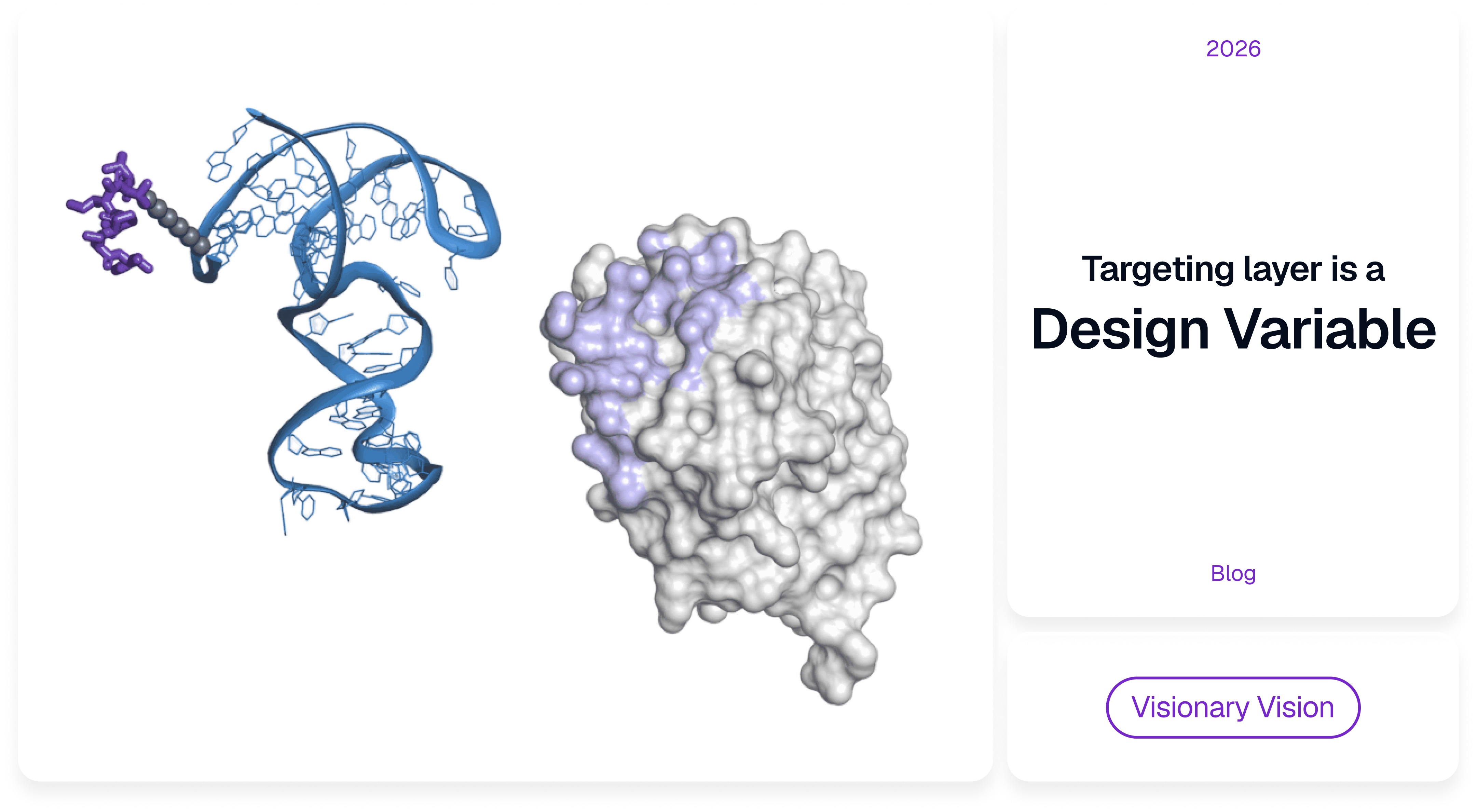
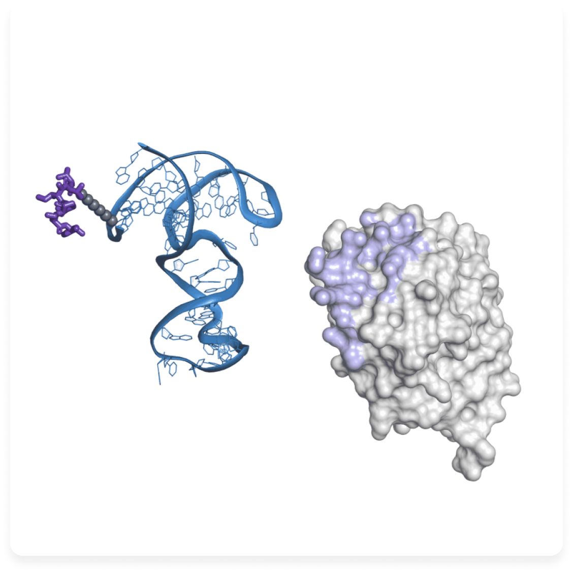
The changing architecture of targeted drugs
A conventional small molecule has to do everything at once. One chemical structure supplies potency, solubility, stability, permeability, distribution and clearance and those properties are entangled: a modification that raises tumor exposure usually raises exposure to healthy organs as well and a longer half-life can improve efficacy while narrowing the therapeutic window.
Targeted delivery split the jobs across components. If one part decides where a construct binds and another supplies the pharmacological effect, a potent compound no longer has to provide its own selectivity. That mattered most for cytotoxics, where potency was never the shortage. The problem was separation – enough distance between tumor exposure and systemic toxicity to make a dose worth giving.
Monoclonal antibodies made the split practical. The hybridoma method Köhler and Milstein published in 19751 turned recognition of a chosen molecular surface into something reproducible and manufacturable. Antibody-drug conjugates then made the architecture explicit: the antibody recognises, the payload acts, the linker decides when the two come apart.
Gemtuzumab ozogamicin demonstrated both halves of the lesson. Accelerated FDA approval in 2000, withdrawal in 2010 after a confirmatory trial found no survival benefit and more fatal induction toxicity, return in 2017 at a fractionated dose. Same molecule, different schedule. Dividing a drug into functional modules does not make the modules independent; the behaviour of the finished construct still comes out of how the parts interact.
Targeting solved recognition before it solved delivery
The intuitive model of targeted delivery is geographical: a ligand recognises an antigen enriched on diseased cells, carries a payload to that molecular address and raises the fraction of drug acting where it should. The address is one point on a much longer route.
A construct administered into the bloodstream has to stay intact long enough to reach the relevant tissue, leave the vascular compartment and cross an extracellular environment that is neither homogeneous nor well mixed. In solid tumors, irregular vasculature, patchy permeability, dense matrix and raised interstitial pressure all shape how macromolecules spread after extravasation. Then it begins meeting target-positive cells and receptor density and binding kinetics decide whether it travels deeper or is captured close to where it arrived.
That is the point at which stronger binding stops paying off. High affinity improves retention at the target, but when binding is fast relative to diffusion, the first accessible cells take up a large share of the incoming dose. Mechanistic penetration models describe the result as a binding-site barrier: steep concentration gradients, with cells far from a vessel exposed to much less drug than a conventional binding assay would predict2.
Selection continues after the receptor is engaged. Surface proteins differ in how fast they internalise, whether they recycle to the membrane and which compartments they enter. Different binding sites on the same receptor can send a construct down different routes. For a delivery system, an epitope is not merely somewhere affinity can be achieved. It can set the rate of internalization and the intracellular fate of whatever is bound to it.
How much that matters depends on the cargo. A small cytotoxin released by lysosomal processing usually survives cleavage and diffuses to its intracellular target. RNA, proteins and gene-editing systems need a narrower route, because entry into the cell is worthless if the cargo stays trapped in an endosome or is degraded before reaching the cytoplasm or nucleus.
So the relevant target for delivery is more specific than a receptor name. It is an accessible molecular surface plus a post-binding route the cargo can survive. Two ligands against the same receptor can match on affinity and still behave as different delivery agents.
None of this was visible while molecular recognition was itself the dominant uncertainty. ADCs made it visible: once a drug can find the target cell reliably, distribution, internalization and intracellular processing become the targeting problem.
Recognition is not physically neutral
An antibody delivers specificity inside a substantial molecular structure. An IgG runs about 150 kDa, with a characteristic geometry, defined valency and pharmacokinetics that can hold systemic exposure for days or weeks. Those properties are why antibodies work as drugs: the protein scaffold supports high-affinity recognition, slow clearance sustains target exposure and decades of development have produced mature manufacturing and conjugation chemistry.
Put one into a delivery system and recognition arrives with all of it. The construct acquires an antigen-binding surface and also an antibody-sized scaffold, antibody geometry and antibody circulation times.
Often that is exactly what the drug wants. Slow clearance gives repeated chances at the target; a large stable scaffold supports complex therapeutic formats. Whether these are advantages depends entirely on the drug being built, not on the class in the abstract.
Which is what makes other recognition scaffolds worth examining. Short DNA or RNA sequences fold into structures that recognise defined molecular surfaces and they differ from antibodies in size, chemistry and route of manufacture. A 40-nucleotide aptamer weighs roughly 12–13 kDa3 and functional groups can be placed at chosen positions during chemical synthesis.
An aptamer is not antibody recognition in a smaller package. It starts from different physics and different chemistry, so properties that come bundled with an antibody can instead be introduced deliberately as the rest of the construct is assembled.
Size and clearance have to be considered together
Size is the obvious contrast. A compact aptamer wired directly to a small-molecule payload stays well below ADC mass, which affects diffusion through tissue and removes some of the physical barriers that come with large protein conjugates. Display the same aptamer on a liposome, polymer particle or large nucleic-acid scaffold and the contrast disappears, because the carrier now sets hydrodynamic size, charge and biodistribution.
Smallness by itself is therefore not the asset. The asset is being able to keep the finished drug compact when payload and delivery strategy allow it.
The smaller starting point immediately drags in another variable. Short nucleic-acid constructs are filtered rapidly by the kidney, while antibodies stay in circulation far longer. For many applications rapid clearance is a real limitation: a construct that disappears before enough material reaches the target cannot deliver an effective dose.
But systemic persistence is only worth having while it is still producing uptake. Once part of the dose is inside the target tissue, the remainder keeps circulating past healthy organs. With very potent payloads that residual exposure becomes a meaningful component of the toxicity.
The relationship that matters is between the rate at which the target acquires drug and the rate at which unproductive material leaves the systemic compartment.
A 2025 conjugate of the PTK7 aptamer Sgc8c and monomethyl auristatin E shows the arrangement concretely4. The finished construct weighed about 14.1 kDa and retained PTK7 binding after conjugation. It accumulated in mouse tumors and produced regression and drug-related material persisted in tumor tissue while normal-organ levels declined. In rats, more than three quarters of measured MMAE-related material was recovered in urine and faeces within 24 hours. The work extended to pharmacokinetic and toxicokinetic evaluation in cynomolgus monkeys, though the antitumor efficacy data remain preclinical.
Fast clearance on its own would be unremarkable. What the study shows is two clocks running at different rates – the drug leaves the blood while material that has already undergone productive uptake stays at the target.
Such a profile would not suit every receptor or payload. Make uptake slower and clearance strips the construct out before the tissue has seen enough. Circulation can be extended by increasing effective size, adding lipid or protein-binding groups, or moving to a larger carrier3 and each of those changes more than one thing at a time: larger size cuts renal filtration and slows tissue penetration, hydrophobic groups shift serum interactions and organ distribution.
Size and half-life, then, are properties of a particular construct rather than of a ligand class.
Chemical definition makes attachment a design variable
The architecture changes again when the payload goes on. Diagrams of targeted drugs show binder, linker and payload as clean functional blocks, but conjugation produces a new molecule whose behaviour does not follow from the properties of the isolated components.
ADC development established this the hard way. Drug-to-antibody ratio, attachment position and linker chemistry all shift solubility, stability, pharmacokinetics and toxicity, which is why controlled and site-specific conjugation has become standard practice.
Aptamers meet the same problem from a synthetic nucleotide scaffold. Functional handles go in at defined positions, so related constructs can be prepared with different attachment sites, spacer lengths, stabilising modifications or additional binding elements. This does not make conjugation biologically neutral. It makes variation cheap to specify.
The effects are real. A payload attached near a structurally important region can weaken binding or disturb folding, while the same payload elsewhere does almost nothing. Hydrophobic cargo changes solubility and can promote aggregation. Modifications introduced to resist nucleases can alter the conformation the target requires. Measurements made on an unconjugated aptamer describe an intermediate component, not the finished drug.
Multivalent systems raise the stakes. Extra copies of a targeting ligand increase avidity and the distance and orientation between them also affect receptor clustering, internalization and local retention. Experiments with DNA nanostructures have shown that constructs of similar molecular composition are taken up differently when the geometry of aptamer presentation changes5.
Geometry becomes part of the mechanism there rather than a formulation detail. The targeting layer can be specified by which molecular surface it recognises and by how several recognition events are arranged in space.
Sequence-level synthesis is what makes that tractable. Attachment position, spacing and valency can be varied systematically and compared experimentally instead of being accepted as fixed features of the targeting format.
The payload defines the useful route
As therapeutic cargoes diversify, the demands placed on the targeting layer get more specific. A cytotoxic small molecule, an RNA therapeutic, a protein and a gene-editing system all benefit from selective cellular recognition. They do not need the same intracellular route.
For a payload such as MMAE, internalization followed by lysosomal processing works, because cleavage frees a small molecule capable of reaching its target from there. RNA is the opposite case: endocytosis delivers it to the correct cell and most of the material stays in compartments it cannot leave. Proteins and gene-editing components add further requirements for protection, release and intracellular localisation.
The preferred receptor and epitope therefore depend partly on the cargo. A target that internalises quickly into lysosomes is attractive for one payload and unhelpful for another. The amount of material required per cell decides something else – whether a compact direct conjugate is realistic at all, or whether a larger carrier is unavoidable.
Which is why aptamer-based delivery is not a single format. A direct aptamer–small-molecule conjugate and an aptamer displayed on a lipid nanoparticle can share a recognition molecule and have very little else in common pharmacologically. In the direct conjugate the aptamer largely sets size, clearance and tissue distribution; on a larger carrier it recognises the receptor while the carrier determines most other physical properties4,6.
Once payload and carrier are specified, the requirements for a targeting ligand get sharper. It may need to engage a particular surface epitope, internalise at a useful rate, tolerate specific chemical modifications and retain its fold after conjugation. Affinity is still necessary. It is no longer sufficient.
For aptamers this connection is unusually direct, because sequence, structure and chemistry are linked. Selection conditions influence which folds are enriched, while salts, serum proteins, nucleases and chemical modifications can change those folds afterwards. A ligand selected against purified protein under convenient laboratory conditions can behave differently in the chemically modified construct that is eventually dosed.
Structural and computational methods can move some of these constraints earlier in the process. Where the epitope and the drug format are already known, candidate structures can be assessed against surface accessibility, planned modifications and attachment positions before anything is synthesised. They do not predict tissue penetration or intracellular pharmacology and they do not replace experimental validation, which remain the established route wherever a target permits them. What they can do is narrow the field for targets where selection is difficult or impractical and remove candidates incompatible with the intended architecture before bench work begins.
The practical difference is one of ordering. The requirements of the drug can inform which binder is developed, rather than arriving after one has already been found.
Comparing architectures rather than ligand classes
The standard antibody-versus-aptamer comparison runs on class properties: antibodies larger and clinically established, aptamers smaller, chemically synthesised and easier to modify. Useful for orientation, poor as a guide to development, because the choice is almost never between two isolated ligand classes.
The comparison that matters is between complete therapeutic architectures built to perform the same biological task.
An antibody is the right targeting layer when its long systemic exposure, protein scaffold and established engineering framework fit that task. An aptamer becomes interesting when changing the recognition scaffold changes something the finished drug depends on: keeping a conjugate compact, defining attachment geometry, controlling valency, engaging a particular epitope or working in a pharmacokinetic regime where uptake is followed by relatively rapid removal of the unbound fraction.
This also changes how ligand discovery connects to drug design. The selection methods introduced in 1990 were built to pull binders out of enormous libraries and they remain the workhorse of aptamer development. But "bind this protein" is a broad specification for a delivery construct. A useful ligand may need a particular epitope, a defined attachment chemistry and a post-binding route the cargo can survive – requirements that can be written down before the search begins rather than discovered after it.
From targeted molecules to designed systems
Each step in this history moved a function out of the active molecule. Monoclonal antibodies made molecular recognition a therapeutic property in its own right. ADCs separated recognition from cytotoxic activity and put a linker in charge of the connection. The drugs that followed showed that modularity could widen the usable range of potent payloads and also that the physical properties of the recognition layer remain part of the pharmacology of the complete construct.
Aptamers extend the same architecture from a different starting point. Because the recognition component can begin as a compact, chemically specified nucleic-acid structure, its size, attachment chemistry, valency and integration with other molecular elements can be arranged differently from an antibody-based system.
Whether that difference is worth anything depends on the therapeutic problem. Some drugs want the long circulation and mature engineering of an antibody and should keep them. Others are limited by scaffold size, clearance kinetics or control over molecular geometry. As payloads expand from cytotoxic small molecules to RNA, proteins and gene-editing systems, one targeting architecture fitting every delivery route looks increasingly unlikely.
What is changing is the status of the targeting layer. It has become a design variable inside the drug rather than a fixed component chosen in advance.
That reframes the question asked at the start of a development programme. Not only which molecule binds the right target, but what kind of recognition event the finished therapeutic actually requires: which surface should be engaged, how quickly the construct should internalise, where it should traffic, how long it should remain in circulation and how its physical architecture interacts with the payload.
Aptamers matter where the answers point to a recognition layer that has to be built differently.
References
- Köhler G., Milstein C. Continuous cultures of fused cells secreting antibody of predefined specificity. Nature. 1975;256:495–497. DOI: 10.1038/256495a0.
- Vasalou C., Helmlinger G., Gomes B. A Mechanistic Tumor Penetration Model to Guide Antibody Drug Conjugate Design. PLOS ONE. 2015;10:e0118977. DOI: 10.1371/journal.pone.0118977.
- Zahedi et al. Aptamer-based drug delivery systems: advances, challenges and future perspectives. International Journal of Pharmaceutics. 2025. DOI: 10.1016/j.ijpharm.2025.125902.
- Su M. et al. An aptamer-drug conjugate for promising cancer therapy with comprehensive evaluation from rodents to non-human primates. Signal Transduction and Targeted Therapy. 2025;10:316. DOI: 10.1038/s41392-025-02399-1.
- Hu et al. Control of multivalent aptamer recognition and cellular internalization through DNA nanostructure geometry. Journal of the American Chemical Society. 2024. DOI: 10.1021/jacs.3c10704.
- Zhao X. et al. Transferrin Receptor-Targeted Aptamer–Drug Conjugate Overcomes Blood–Brain Barrier for Potent Glioblastoma Therapy. Bioconjugate Chemistry. 2025. DOI: 10.1021/acs.bioconjchem.5c00137.
© 2026 Xelari Inc. All rights reserved.
