
Why change the chemistry of aptamers?
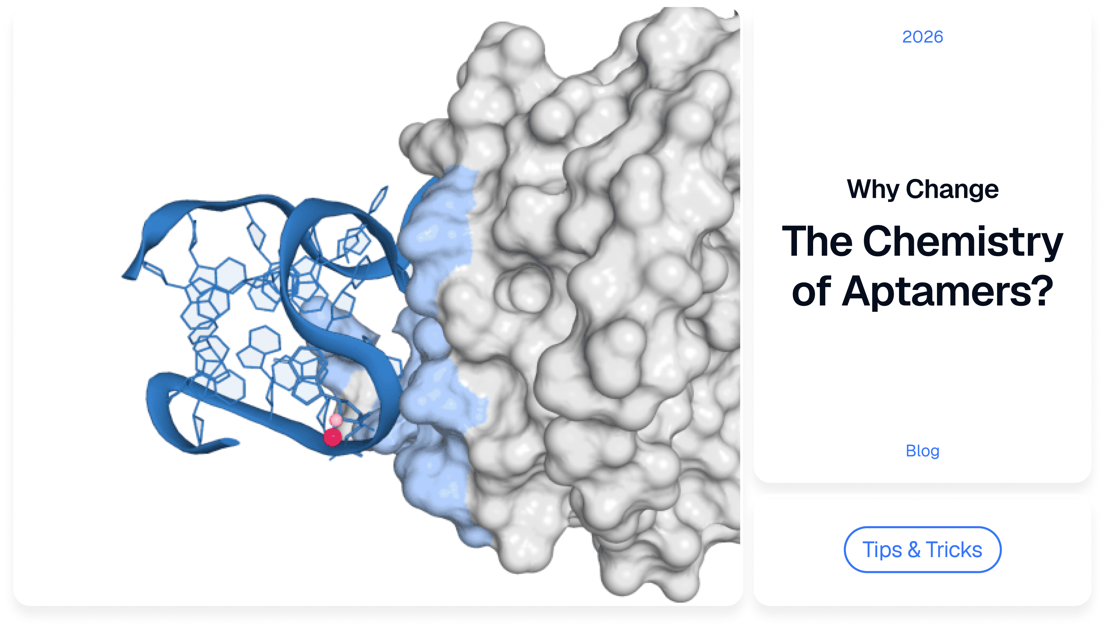
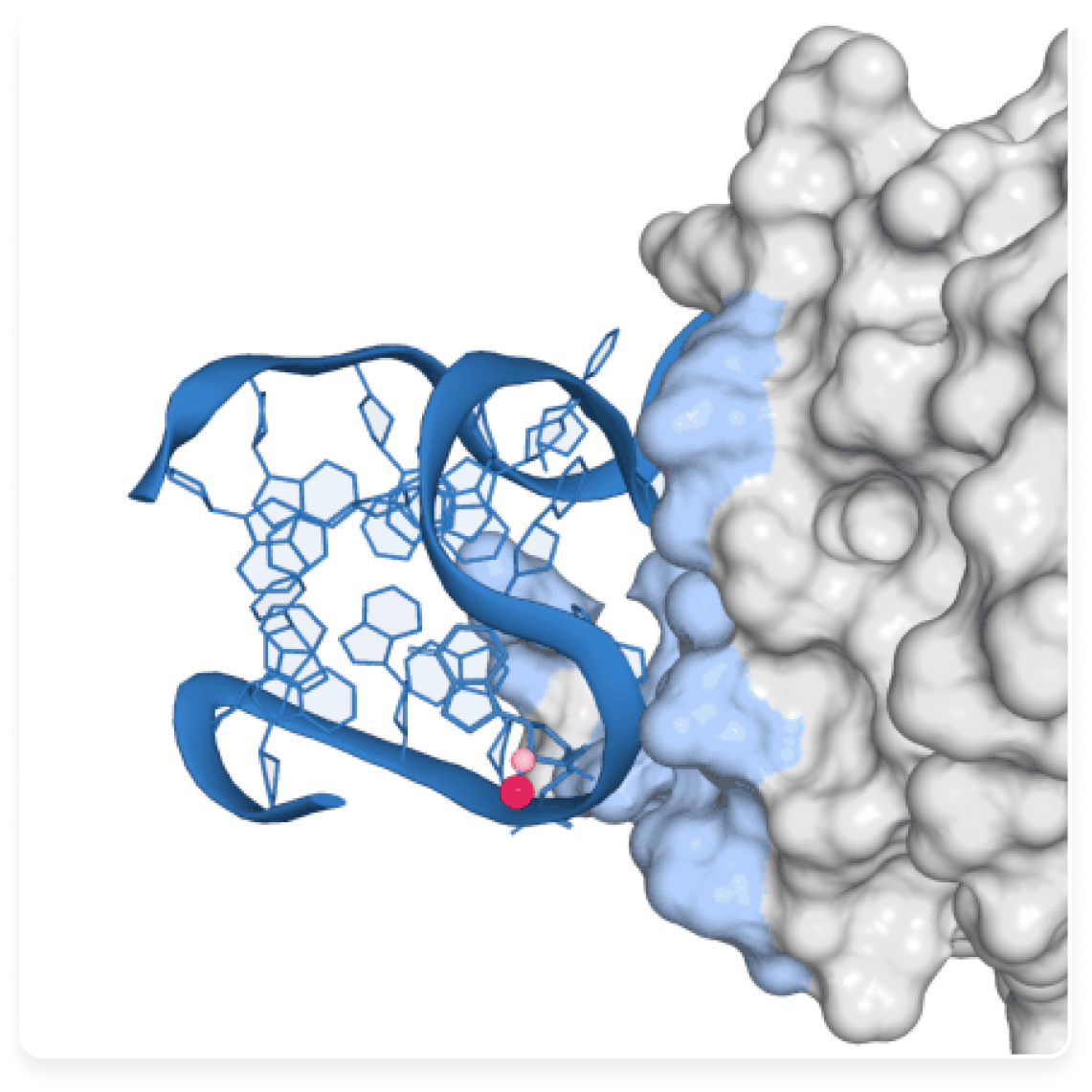
Chemical modifications have a peculiar place in aptamer development. They often come into play only after something has already gone wrong: an aptamer is degraded too quickly in serum, cleared from the bloodstream too rapidly, or loses activity at 37 °C. The usual response is to introduce 2′-OMe residues, protect one end, attach PEG and measure the properties again.
That approach makes sense, but a chemical modification rarely affects only the parameter it was meant to improve. Replacing a single atom or a small functional group can substantially change how long a molecule survives. Modified bases can form contacts with proteins that natural DNA or RNA cannot, while a change introduced to improve stability can alter the geometry of the binding site and, with it, affinity. At the atomic level, every substitution creates a different molecule, with different energetics, dynamics and sometimes a different mode of interaction with its target.
Stability is not one property
An aptamer binds its target because of its three-dimensional structure. Its sequence determines which structures are accessible, but a string of A, G, C and T or U does not bind a target on its own. In solution, stems transiently open and close, loops shift position, bases rotate and the chain moves among conformations that are close in energy. One of these conformations, or several closely related states, is capable of binding the target.
In a biological environment conformational dynamics is only part of the picture. Nucleases cleave nucleic acids. Ionic conditions change. Proteins, lipids and other potential interaction partners surround the molecule. After systemic administration, the small size of an aptamer can lead to rapid renal clearance. If its functional structure is not sufficiently stable at body temperature, a substantial fraction of the molecules may also occupy conformations that bind poorly.
These effects are sometimes grouped under the word stability, but physically they are different phenomena. Nuclease resistance tells us nothing about the rate of renal clearance. A high melting temperature does not guarantee high affinity. Long residence in the bloodstream does not mean that the molecule retains its functional structure throughout that time. If the cause of activity loss is misidentified, chemical optimisation can improve a parameter that was never the main limitation.
2′-F and 2′-OMe can protect RNA and change its structure
One of the most common targets for chemical optimisation of RNA is the sugar moiety of the nucleotide. Ribose carries a hydroxyl group at the 2′ position; replacing it with fluorine gives a 2′-F nucleotide, while a methoxy group gives 2′-O-methyl, or 2′-OMe.
These substitutions can substantially increase resistance to enzymatic degradation. The degree of protection, however, depends both on which positions are modified and on the structure of the chain itself. Fully modifying an aptamer is not automatically the best strategy, because changing the sugar affects more than the interaction with a nuclease: it can alter the preferred geometry of the nucleotide, the flexibility of the chain and the relative positions of the bases.
In a stem, where a nucleotide mainly serves a structural role, a substitution may have little effect on binding. At a tightly packed protein-binding interface, even a small displacement can reorganise local contacts. The same 2′ modification can therefore improve nuclease resistance while reducing affinity. Stability of the modified aptamer should be evaluated alongside binding rather than treated as an independent optimisation target.
Terminal protection addresses a narrower problem. An inverted thymidine at the 3′ end, for example, can prevent some exonucleases from initiating degradation at that terminus, but it does not protect against enzymes that cleave the oligonucleotide internally. The name of a chemical group may suggest how it is likely to act; it does not determine the outcome for a particular aptamer.
Phosphorothioate stereochemistry can change function
Phosphorothioate linkages are a good example of why sequence alone may be insufficient to describe a chemically modified aptamer. In a conventional phosphate backbone, one of the non-bridging oxygen atoms can be replaced by sulfur. Oligonucleotides containing these linkages are often more resistant to nucleases and their interactions with proteins can change as well.
The substitution introduces another feature: the phosphorus centre becomes chiral. A phosphorothioate linkage can therefore exist in two spatial configurations, while standard synthesis commonly produces a mixture of them. In a 2025 study using aptamers and DNAzymes as model systems, stereochemical variants with identical sequences differed in melting temperature by as much as 15 °C and their functional properties differed as well [1].
For natural DNA, sequence can often serve as a sufficiently precise identifier of the molecule. A chemically modified aptamer requires a more complete description. The positions and types of modifications matter and for some chemistries so does stereochemistry. If binding is the property of interest, the functional three-dimensional structure matters as well.
Modified bases expand the chemistry of recognition
Chemical modification is not limited to protecting an aptamer that already exists. It can also expand the set of interactions available for binding the target. Protein surfaces are chemically diverse: amino-acid side chains provide positive and negative charges, hydrogen-bond donors and acceptors, aromatic rings and hydrophobic regions. Natural DNA and RNA have a more limited repertoire of accessible functional groups, particularly large hydrophobic side groups.
Additional chemical groups can be attached directly to the bases. Some SOMAmers use this principle: their modified pyrimidines carry hydrophobic aromatic substituents that can make additional contacts with protein surfaces. In one well-studied example, optimised SOMAmers against interleukin-6 reached a dissociation constant, KD, of about 0.2 nM [2]. A co-crystal structure showed that the added groups participate directly in contacts with the protein [3].
In this case, the modification does more than improve stability or pharmacokinetics. It expands the chemical space available for molecular recognition. Hydrophobicity alone, however, does not confer specificity. The same group that interacts favourably with the intended region of a target protein can also increase nonspecific binding to other proteins. Position therefore matters: what counts is where the additional group points and what molecular environment surrounds it in the complex.
More rigidity does not always improve binding
Another way to change the properties of an aptamer is to restrict the mobility of its chain. In LNA, or locked nucleic acid, the sugar is chemically constrained in a particular conformation. LNA nucleotides often increase the thermal stability of structural elements, which can be useful if the stabilised state resembles the functional conformation of the aptamer.
But binding does not always occur between two structures that are already rigid and fully preorganised. A free aptamer may populate several states at once. When the target appears, the equilibrium between those states can shift and formation of the complex may require local structural rearrangements. If a modification stabilises a favourable geometry, it can reduce the conformational cost of binding; if it locks the aptamer into the wrong state, affinity can fall.
A high melting temperature, Tm, therefore does not by itself show that the molecule has become functionally better. The relevant question is which structure has gained stability and where the modification sits relative to the binding motif.
Mirror-image aptamers resist natural nucleases
Spiegelmers rely on a different principle. They are built from L-nucleotides, the mirror-image counterparts of natural D-nucleotides. Biomolecules are chiral, which means that an enzyme-substrate interaction depends not only on the chemical groups involved but also on how those groups are arranged in space. The geometry of an L-nucleic acid is poorly matched to the recognition and catalytic sites of most natural nucleases, so these molecules are generally degraded much more slowly.
Here, resistance is achieved not by protecting individual bonds but by changing the chirality of the entire chain. Several Spiegelmer candidates have reached clinical studies [4]. The example illustrates a broader point: two aptamers can persist for similar lengths of time in a biological environment while achieving that persistence through different molecular mechanisms and with different accompanying properties.
Nuclease resistance and circulation time are different problems
Even an aptamer that is well protected from nucleases may disappear rapidly from the circulation after systemic administration. Short oligonucleotides are small and the kidneys efficiently clear low-molecular-weight molecules. An aptamer can therefore remain chemically intact while still being removed from the bloodstream.
Protecting the phosphate backbone does not solve this problem. Instead, the effective size of the construct or its interactions with components of blood must be altered. One of the best-known approaches is PEGylation, which increases hydrodynamic size and changes pharmacokinetics. Pegaptanib, the first approved aptamer drug, is a PEGylated anti-VEGF aptamer [5].
Another strategy is to introduce interaction with albumin. Albumin is abundant in plasma and circulates for a long time, so binding to it can change the distribution and persistence of an aptamer in the bloodstream. A hydrophobic or albumin-binding group, however, can also affect interactions with the primary target and with other proteins. A pharmacokinetic module therefore has to be evaluated as part of the complete molecule.
Labels and immobilisation can change binding
ESome modifications are almost automatically treated as purely technical additions. Biotin is used for immobilisation; a fluorophore is used to detect a signal. Because these groups are introduced for experimental reasons, it is easy to treat their effects on the aptamer itself as secondary.
At the molecular level, however, there is no separate category of “technical” groups. Cy5 has a particular size and charge. Biotin is connected to the chain through a linker of a particular length. Once either is attached, the steric bulk and mobility of that region change. If a label is far from the binding interface, its effect may be small; if the end of the chain lies close to the target surface, the label can alter binding.
Immobilisation changes the experimental system as well. A free aptamer can approach a protein from different directions, whereas a surface-tethered aptamer is constrained by the position of the linker and the geometry of the sensor. Binding should therefore be measured again after adding a label or moving to an immobilised format.
The application determines how much modification is useful
There is no universal number of modifications an aptamer needs. The requirements depend on the application. A diagnostic aptamer that operates for several minutes in a prepared sample may have no need for prolonged circulation. For a systemically administered drug, several hours of serum stability are of little value if the molecule is cleared from the bloodstream before it reaches enough target tissue. Local administration creates a different set of requirements.
Even high affinity can mean different things in different settings. Sometimes a slow dissociation rate matters most. In other cases, the rate of complex formation is more important, or the key requirement is retaining binding in the presence of a large excess of background proteins. The practical goal is therefore to achieve properties that are sufficient for the intended environment and mode of use without unnecessarily changing regions that already work.
Structural information can guide chemistry before synthesis
The familiar development sequence is sequential: an aptamer is first obtained using standard nucleotides, its weaknesses are characterised and chemical modifications are introduced afterwards. The advantage is that the binding properties of the starting molecule are already known. The drawback is that every subsequent substitution has the potential to disrupt a structure that originally formed without it.
A useful question is whether some of those choices can be made before a modified candidate is synthesised. If the three-dimensional structure of the target complex is known, a proposed modification can be assessed in structural terms: does the nucleotide contact the protein, is there enough space nearby for a bulkier group, which way does the end of the chain point and which regions mainly serve as structural scaffolds? Computational models can help formulate these hypotheses, but they do not establish that a particular chemistry will work.
The modified molecule still has to be synthesised and measured. Nuclease resistance, conformational or thermal behaviour, target binding and, where relevant, pharmacokinetics are separate readouts. Structural information can reduce arbitrary substitutions and focus the first experimental set; it does not remove the need to evaluate the resulting aptamer as a whole.
References
- Stereochemistry of Phosphorothioate Linkages Impacts the Structure and Binding Affinity of Aptamers and DNAzymes. Molecular Pharmaceutics. 2025. DOI: 10.1021/acs.molpharmaceut.5c00117.
- Gupta S. et al. Chemically Modified DNA Aptamers Bind Interleukin-6 with High Affinity and Inhibit Signaling by Blocking Its Interaction with Interleukin-6 Receptor. Journal of Biological Chemistry. 2014. DOI: 10.1074/jbc.M113.532580.
- Gelinas A.D. et al. Crystal Structure of Interleukin-6 in Complex with a Modified Nucleic Acid Ligand. Journal of Biological Chemistry. 2014. DOI: 10.1074/jbc.M113.532697.
- Vater A., Klussmann S. Turning mirror-image oligonucleotides into drugs: the evolution of Spiegelmer therapeutics. Drug Discovery Today. 2015. DOI: 10.1016/j.drudis.2014.09.004.
- Macugen AMD Study Group. Pegaptanib 1-year systemic safety results from a safety–pharmacokinetic trial in patients with neovascular age-related macular degeneration. Ophthalmology. 2007. DOI: 10.1016/j.ophtha.2007.02.021.
© 2026 Xelari Inc. All rights reserved.
