
Why pH, temperature and ions belong in the design specification
The same aptamer sequence works reliably in one laboratory and produces a weak signal in another. Has something gone wrong with the DNA? Perhaps. But first ask whether the two laboratories have given the molecule the same chemical environment.
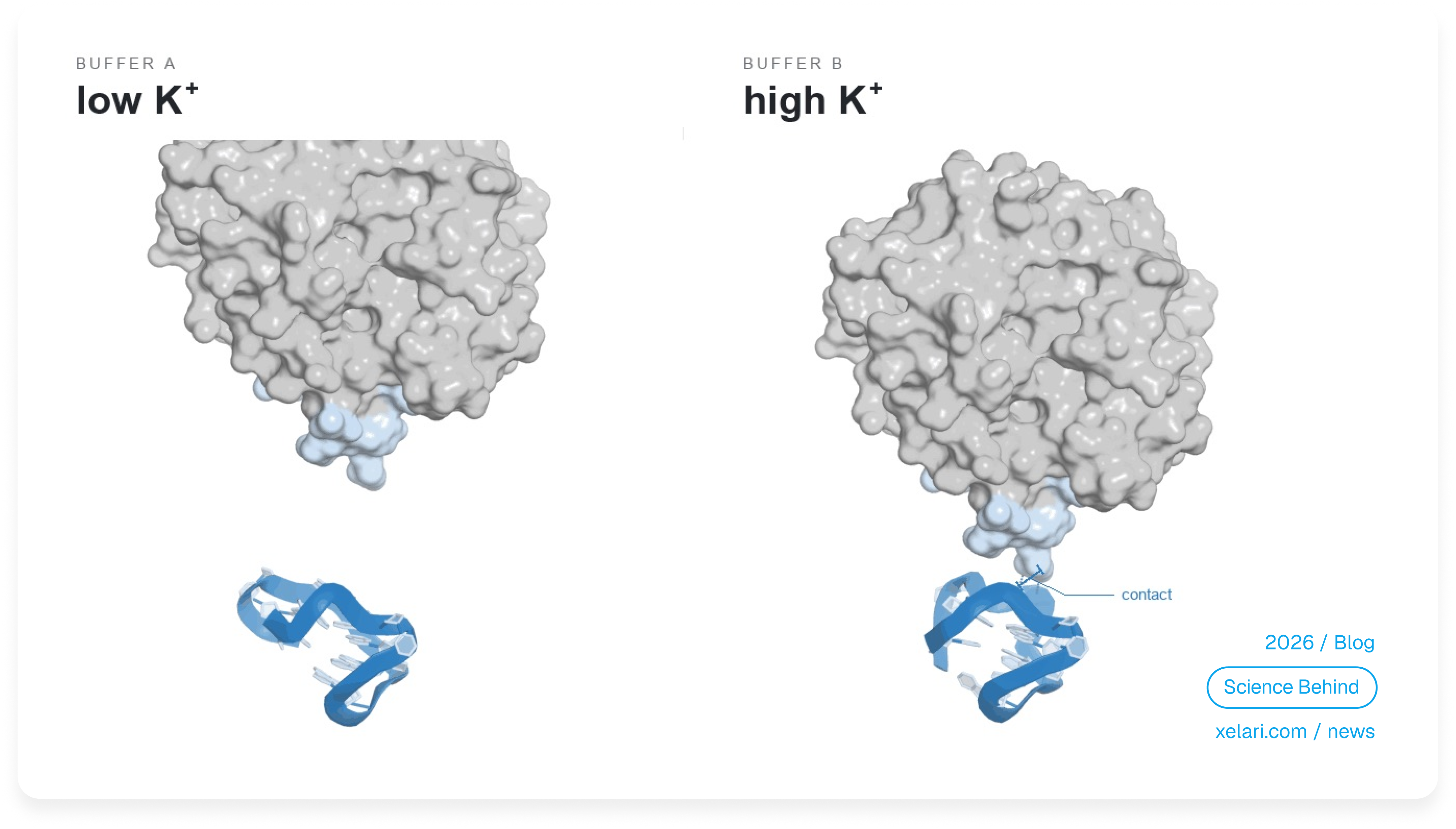
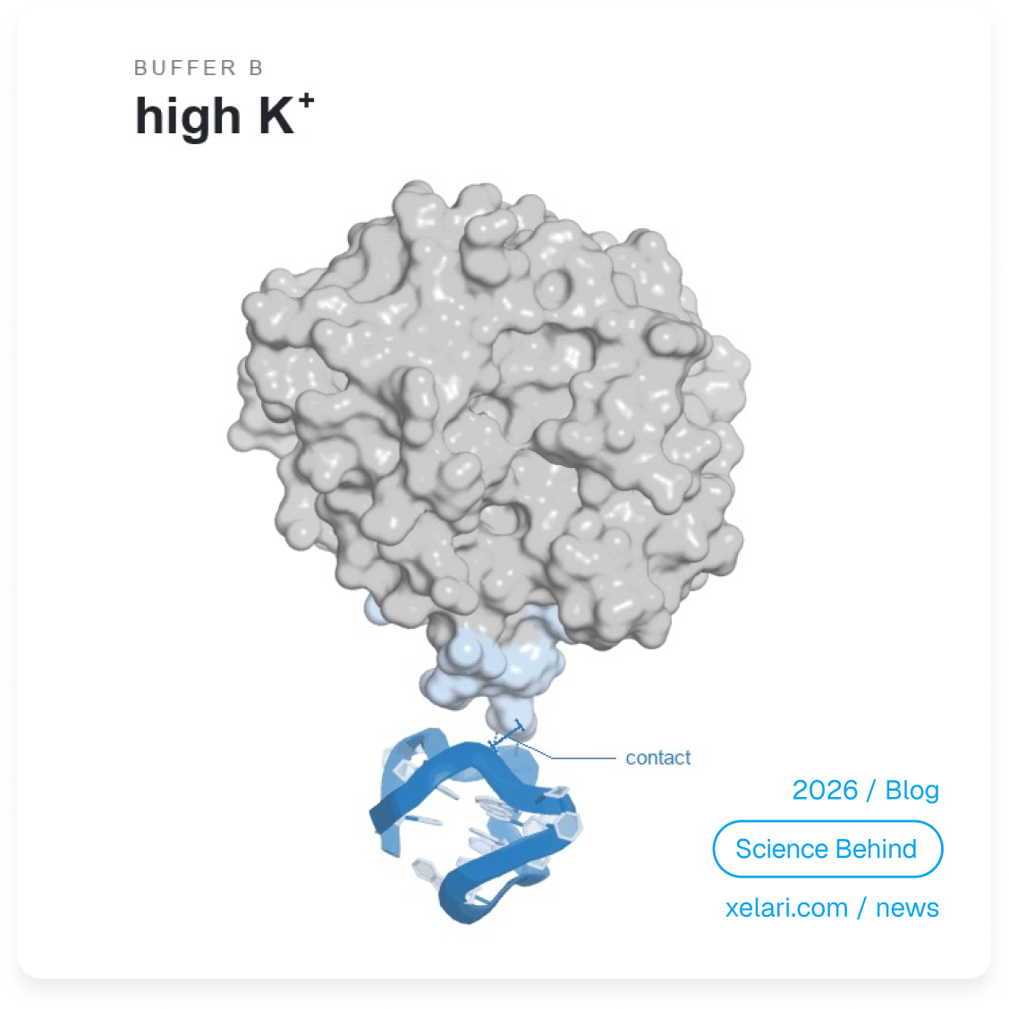
An aptamer is a short strand of DNA or RNA that recognizes a molecular target through its three-dimensional arrangement and chemical contacts. Unlike a rigid manufactured component, it can change shape. Experiments that track individual aptamers show that ions can alter the balance between those shapes without changing a single nucleotide–the building block of the strand. [1] [2]
Sequence defines the possibilities. The environment helps determine which states are populated, which contacts are favorable and whether productive binding can occur. Operating conditions therefore belong in the design specification.
A moving population, not a single pose
Single-stranded DNA and RNA can fold back on themselves. Complementary regions form paired stems; unpaired regions create loops; more distant parts make three-dimensional contacts. Several arrangements may be accessible because different contacts can form, break, or compete. Thermal motion keeps the molecule exploring those possibilities. “Single-stranded” does not mean unstructured and “structured” does not mean motionless. [1] [2]
A conformational ensemble is the collection of accessible shapes and their relative populations. It need not be a chaotic mixture: one shape may dominate while others appear briefly. A useful mental picture is a landscape with several valleys. Each valley represents a family of similar conformations. Changing the environment changes their relative depths and the barriers between them.
At equilibrium, deeper valleys – states with lower free energy, are more populated. Free energy accounts for the molecule together with its interactions with solvent and ions. A condition change may therefore make an existing, rare state common rather than create a wholly new fold. If transitions are slow, the population observed during an assay can also depend on how the sample was prepared. [3] [4]
Function depends on binding-competent states: arrangements from which the target can engage productively. In an ATP-binding DNA aptamer, a bound-like shape appeared even without ATP. At low salt it was short-lived; ATP stabilized it. This supports conformational selection, in which binding favors a suitable state already present. Further adjustment after contact–induced fit can also occur. These are compatible mechanisms, not competing universal explanations. [2] [5]
Salt acts on both the fold and the binding interface
Near neutral pH, the phosphate backbone of DNA or RNA is negatively charged. Folding brings some of these charges closer together, creating repulsion. Positive ions accumulate preferentially near the strand, while negative ions are relatively depleted. This dynamic ion atmosphere screens electrostatic interactions: it reduces their effective range and strength without removing the backbone’s chemical charge. [6]
Ionic strength summarizes the concentrations and charges of all ions in solution:
Here, cᵢ is the concentration of ion type i and zᵢ is its charge number. Anions count too. Thus, 100 mM NaCl has an ionic strength of 100 mM, whereas 1 mM MgCl2 contributes 3 mM. Equal calculated ionic strength, however, does not make two solutions chemically equivalent.
Screening can make compact folding less costly. At the target interface, its effect depends on the local charges: salt can weaken helpful attraction between the negative backbone and a positive protein patch, or ease an approach hindered by repulsion. Water and ions also redistribute when the complex forms. The net outcome reflects both folding and association, not a simple “more salt is better” rule. [6] [7]
A microbead study illustrates this distinction. Increasing NaCl generally reduced aptamer–protein binding signals, but one system showed a maximum at an intermediate concentration. The measurement demonstrates a condition-dependent response; it does not establish a universal salt optimum or prove which molecular step changed. [8]
Na+ and K+: similar screening, different local chemistry
Sodium and potassium both carry one positive charge. Equal concentrations of NaCl and KCl therefore produce equal calculated ionic strength. Yet Na+ and K+ differ in size and hydration–the way water surrounds them. They can occupy the molecular environment differently and stabilize particular contacts to different extents. [9]
Such specificity is architecture-dependent. A comparison of RNA structures found that simple hairpins discriminated little among monovalent cations, whereas some contacts joining distant RNA regions were more sensitive. The ligand-binding domain of an adenine riboswitch, a natural RNA regulator, also showed ion-dependent behavior. These RNA models illustrate physical mechanisms; they do not establish the response of every designed DNA or RNA aptamer. [9]
For design, the implication is straightforward: record Na+ and K+ separately rather than replacing them with “physiological salt.” Their ratio matters when the particular structure or interface is ion-sensitive; neither interchangeability nor a mandatory advantage of one ion should be assumed.
Why magnesium can be decisive or barely noticeable
Mg2+ carries two positive charges and interacts strongly with regions where negative charge is concentrated. One contribution is diffuse stabilization: magnesium in the surrounding ion atmosphere can favor a folded state without occupying a unique structural “socket.” Experiments on a compact RNA model demonstrated this mechanism directly. [3]
A second contribution is local coordination. Magnesium may interact with RNA through its surrounding water molecules, or directly contact suitable atoms after replacing some water in its coordination shell. These interactions depend on geometry and hydration, not merely charge. Describing Mg2+ as a clip joining two phosphates misses much of this chemistry. [10]
Why do sequences respond differently? A fold that gathers several charged regions into a tight junction may benefit greatly from magnesium. Another may already be adequately organized in a monovalent-ion background. Crucially, magnesium changes the ensemble only insofar as it stabilizes competing states differently. Stabilizing a functional and a nonfunctional state equally would leave their equilibrium ratio unchanged. [3] [10]
A weak response over the tested range can also mean that an important magnesium-dependent transition has already occurred. It does not prove that magnesium never mattered. Conversely, some RNA architectures can fold and bind ligand without Mg2+ when monovalent-ion conditions are suitable. [9] [11]
For a tetracycline-binding RNA aptamer, one study found that at least about 0.5 mM Mg2+ was needed to form a preorganized, binding-competent pocket under its experimental conditions. Binding then triggered further rearrangements. The number is evidence for a mechanism in that system–not a recommended concentration for other aptamers. [5]
Finally, added magnesium is not necessarily free magnesium. Other components may bind it, for example through chelation. Yet “bound” does not always mean unavailable: weakly chelated magnesium supported folding in RNA-model experiments. Report total magnesium, distinguish any estimate of free Mg2+ and retain the full mixture composition. A concentration alone cannot describe all of its chemical forms. [11]
pH changes the chemistry of recognition
Changing pH changes the likelihood that a chemical group carries a proton. This can alter both its charge and its ability to donate or accept a hydrogen bond. A binding pocket may keep roughly the same outline while its pattern of favorable contacts changes. A group is especially sensitive when its pKₐ – the pH at which its protonated and unprotonated forms are equally populated is near the working pH. [12]
Not every nucleotide switches charge around neutral pH. Many isolated base groups have pKₐ values well outside that range, but their local molecular environment can shift them. In a natural riboswitch, researchers identified a protonated adenine that enabled recognition of an alternative ligand. This is a specific example of protonation changing molecular recognition, not a universal property of adenines in aptamers. [12]
The target matters too. Protein side chains and ionizable groups on small molecules respond to pH. A change in binding can therefore originate in the aptamer, the target, or both. In the microbead study, acidic conditions increased binding while reducing specificity: a larger signal was not necessarily better recognition. [8]
Protonation can also strengthen the wrong fold. Engineered ATP-aptamer switches used pH-sensitive contacts in a competing stem to alter access to the binding state. Stabilizing that competing structure suppressed target binding. This supplies a concrete warning against treating structural stability as a synonym for function. [13]
Temperature changes populations and the clock
Temperature does more than eventually melt a structure. It changes the balance between stabilizing interactions and the number of configurations available to the strand, water and ions. In thermodynamic shorthand, ΔG = ΔH − TΔS: ΔG is the free-energy change, ΔH the enthalpy change associated with interactions, ΔS the entropy change reflecting accessible configurations and T the absolute temperature in kelvin. [14]
Consequently, warming need not affect every aptamer in the same direction. It may weaken a useful stem, but it may also release a competing structure that blocks recognition. Binding adds its own temperature dependence. A large study of DNA aptamers for small molecules found varied enthalpic and entropic contributions to recognition; its results should not be generalized automatically to protein targets or RNA aptamers. [14]
Temperature also changes how quickly transitions occur. A molecule that can reach a productive state eventually may not reach it within a short assay. In the malachite-green RNA aptamer, the apparent binding-active population depended on temperature, magnesium concentration and the time allowed between ligand additions. Higher temperature or lower magnesium made more RNA accessible to binding in particular experimental regimes. [4]
This separates two questions: which states are favored and how quickly can the system reach them? Prefolding, cooling and incubation history matter when slow transitions prevent the sample from equilibrating before measurement.
The most stable fold is not necessarily the most useful
Consider two states. One exposes the recognition pocket. The other traps essential bases inside a competing stem. Stabilizing the second state makes the strand more consistently folded but increases the cost of reaching the binding state. Preorganization helps only when it organizes the right contacts.
For a switching sensor, useful function requires more than a binding-ready pocket: target engagement must produce a measurable change. A structure held too firmly in one state can reduce that response. Engineered aptamer switches demonstrate that the balance between competing structures affects sensitivity, background and response kinetics. A useful design must leave access to the transitions its function requires. [15]
This explains why optimal folding conditions and optimal working conditions can differ. A preparation protocol may favor a reproducible starting population; the assay must support recognition, selectivity and an appropriate response time. A melting transition or a compact structural signal alone cannot establish all three.
A new buffer or a new assay - is a new test
A buffer name is not a complete specification. Matching pH does not match ionic composition and matching added MgCl2 does not match free Mg2+. Other solutes can alter ion availability, while the target encounters the same changed environment as the aptamer. [11]
Assay format adds another layer. Attaching an aptamer to a surface restricts its motion and places it near neighboring strands. Attachment geometry, crowding and target access can change productive binding. In aptamer microarrays, target capture passed through a maximum as surface density increased: simply adding more aptamers was not an unlimited improvement. [16]
Results obtained in one buffer are therefore useful starting evidence, not an unconditional prediction for another experimental system. Robustness is something to establish, not something an unchanged sequence guarantees. Record the complete formulation, final concentrations, preparation history, incubation time and surface format so that disagreements can be investigated.
Start the Xelari design brief at the point of use
Begin with the final assay or application, not with a convenient folding recipe. Which conditions are fixed by the sample or device? Which can genuinely be adjusted? Define those constraints before choosing sequences.
When working with Xelari, specify the actual operating conditions of the final system: temperature, pH, Na+, K+ and Mg2+. These should describe the mixture in which the aptamer encounters its target, after sample addition and dilution–not merely the stock solution.
| Parameter | What the design specification should describe |
|---|---|
| Temperature | The temperature of target interaction and readout, not a preliminary heating step. |
| pH | The pH of the complete working mixture at its operating temperature. |
| Na+ | The final sodium concentration from all sources, including buffer salts and pH adjustment. |
| K+ | The final potassium concentration, recorded separately from sodium. |
| Mg2+ | The final specified magnesium concentration, explicitly distinguishing total content from any estimate of free Mg2+. Record magnesium-binding additives. |
Keep the full buffer recipe, sample fraction, surface chemistry and assay duration alongside these inputs. Where conditions vary, document the intended operating range as well as a nominal point. The five parameters are an essential description of the task, not an exhaustive representation of the experimental system.
A worked example: design for the final mixture
Suppose an assay is intended to operate at 37 °C and pH 7.4. The aptamer is first folded separately in a preparation containing 5 mM MgCl2. One volume is then mixed with four volumes of sample and other reagents. These are illustrative conditions, not a buffer recommendation.
The preparation contributes 1 mM total magnesium to the final mixture, not 5 mM. Magnesium from the sample and other reagents must be added to that accounting; their magnesium-binding components affect how much remains free. Calculate final Na+ and K+ from all inputs in the same way and verify pH in the completed mixture rather than averaging the starting pH values.
The Xelari brief should describe 37 °C, the verified working pH and those final ion conditions. Record the prefolding recipe separately. Then test whether the aptamer retains useful function after the transfer and over the assay’s actual measurement window.
Validate the fold, the binding and the function
Test realistic variations around the intended operating point, including selected combinations rather than only one parameter at a time. Distinguish structural change, target recognition and assay performance. A distance-sensitive fluorescence measurement can probe folding; a binding experiment can assess target interaction; the final assay must establish signal, background, speed and reproducibility. [1] [8] [15]
Affinity measurements, including the dissociation constant Kd, are one way to show that conditions change system behavior. They should sit alongside evidence about the binding-accessible population, kinetics and selectivity–not replace it. Include target-free and non-target controls to distinguish improved recognition from a change in background. [4] [15]
For an application, a tolerant operating range may be more valuable than a sharp maximum under one finely tuned condition. The objective is reproducible function where the molecule will actually be used.
Back to the two laboratories
The first question is no longer just “Did we use the same sequence?” It is “Did we use the same final chemistry, temperature, preparation history and assay format?” Different answers provide concrete explanations to test–not proof that the sequence itself is defective.
An aptamer’s functional form is a property of the system it inhabits. Design therefore begins with sequence, environment, target and application together.
References
- Taylor, J. N., et al. (2008). Dynamics of an anti-VEGF DNA aptamer: a single-molecule study. Biochemical and Biophysical Research Communications, 373(2), 213–218. doi: 10.1016/j.bbrc.2008.05.191
- Xia, T., Yuan, J., & Fang, X. (2013). Conformational Dynamics of an ATP-Binding DNA Aptamer: A Single-Molecule Study. The Journal of Physical Chemistry B, 117(48), 14994–15003. doi: 10.1021/jp4099667
- Soto, A. M., Misra, V., & Draper, D. E. (2007). Tertiary Structure of an RNA Pseudoknot Is Stabilized by “Diffuse” Mg2+ Ions. Biochemistry, 46(11), 2973–2983. doi: 10.1021/bi0616753
- Sokoloski, J. E., Dombrowski, S. E., & Bevilacqua, P. C. (2012). Thermodynamics of Ligand Binding to a Heterogeneous RNA Population in the Malachite Green Aptamer. Biochemistry, 51(1), 565–572. doi: 10.1021/bi201642p
- Kaiser, C., et al. (2023). Magnesium Ion-Driven Folding and Conformational Switching Kinetics of Tetracycline Binding Aptamer: Implications for in vivo Riboswitch Engineering. Journal of Molecular Biology, 435(20), 168253. doi: 10.1016/j.jmb.2023.168253
- Bai, Y., et al. (2007). Quantitative and Comprehensive Decomposition of the Ion Atmosphere around Nucleic Acids. Journal of the American Chemical Society, 129(48), 14981–14988. doi: 10.1021/ja075020g
- Kuo, T.-C., et al. (2013). Salt bridge exchange binding mechanism between streptavidin and its DNA aptamer–thermodynamics and spectroscopic evidences. Journal of Molecular Recognition, 26(3), 149–159. doi: 10.1002/jmr.2260
- Schmidt, C., et al. (2022). A multiparametric fluorescence assay for screening aptamer–protein interactions based on microbeads. Scientific Reports, 12, 2961. doi: 10.1038/s41598-022-06817-0
- Lambert, D., et al. (2009). The Influence of Monovalent Cation Size on the Stability of RNA Tertiary Structures. Journal of Molecular Biology, 390(4), 791–804. doi: 10.1016/j.jmb.2009.04.083
- Leonarski, F., et al. (2025). Principles of ion binding to RNA inferred from the analysis of a 1.55 Å resolution bacterial ribosome structure – Part I: Mg2+. Nucleic Acids Research, 53(1), gkae1148. doi: 10.1093/nar/gkae1148
- Yamagami, R., et al. (2018). Cellular conditions of weakly chelated magnesium ions strongly promote RNA stability and catalysis. Nature Communications, 9, 2149. doi: 10.1038/s41467-018-04415-1
- Keller, H., et al. (2018). Adenine protonation enables cyclic-di-GMP binding to cyclic-GAMP sensing riboswitches. RNA, 24(10), 1390–1402. doi: 10.1261/rna.067470.118
- Thompson, I. A. P., et al. (2020). Rational design of aptamer switches with programmable pH response. Nature Communications, 11, 2946. doi: 10.1038/s41467-020-16808-2
- Alkhamis, O., et al. (2025). Exploring the relationship between aptamer binding thermodynamics, affinity and specificity. Nucleic Acids Research, 53(6), gkaf219. doi: 10.1093/nar/gkaf219
- Wilson, B. D., et al. (2019). Independent control of the thermodynamic and kinetic properties of aptamer switches. Nature Communications, 10, 5079. doi: 10.1038/s41467-019-13137-x
- Simon, L., Bognár, Z., & Gyurcsányi, R. E. (2020). Finding the Optimal Surface Density of Aptamer Monolayers by SPR Imaging Detection-based Aptamer Microarrays. Electroanalysis, 32(4), 851–858. doi: 10.1002/elan.201900736
© 2026 Xelari Inc. All rights reserved.
